Skin Conditions in Dogs
Compound evidence detail1 SCR / 2 parts
- Documentedthe parasite-then-food-then-atopy diagnostic hierarchy that rules out parasitic disease and cutaneous adverse food reaction before working up canine atopic dermatitis; the multi-modal long-term CAD treatment combining allergen avoidance, antipruritic therapy, skin-barrier support, and cytology-guided antimicrobial control of secondary infection; and Golden Retriever elevated CAD predisposition
- Documentedthe high frequency of secondary Staphylococcus pseudintermedius bacterial and Malassezia pachydermatis yeast overgrowth among atopic-dermatitis patients as the clinical rule rather than the exception
Skin disease is one of the most common reasons dogs visit veterinarians, and Golden Retrievers are one of the breeds families most readily associate with itch, recurrent hot spots, ear trouble, and chronic allergy management. That reputation exists for a reason. Goldens are not defined by skin disease, but they are common enough participants in the allergy-and-inflammation world that families benefit from a practical framework instead of a rotating series of creams, wipes, and guesses. Mixed Evidence
What It Means
The Main Skin Categories Families See
Most day-to-day canine dermatology belongs to a few broad buckets atopic dermatitis; secondary bacterial or yeast overgrowth; hot spots; food-responsive skin disease; flea allergy dermatitis; contact reactions; and less common parasitic or autoimmune disease. Documented
These often overlap. A dog does not only have "allergy" or "infection." More commonly, the allergic dog develops secondary infection because the skin barrier has been inflamed and damaged.
Atopic Dermatitis
Atopic dermatitis is the center of gravity in chronic canine skin disease. These dogs are reacting to environmental allergens, and the skin barrier becomes inflamed, itchy, and vulnerable. The classic sites include feet, face, ears, armpits, groin, and belly.
Goldens are one of the breeds families and veterinarians frequently place in this conversation. The practical importance is not the label alone. It is that long-term control usually depends on addressing the allergic biology, not only treating the latest flare.
Why Infection Is So Often Secondary
When a dog itches, chews, scratches, and licks chronically, the skin barrier weakens. Mixed Evidence That creates an opportunity for bacterial pyoderma or Malassezia yeast overgrowth. The family then notices odor, greasy skin, redness, crusting, pustules, or brown staining and quite reasonably thinks "infection."
They are not wrong. They are just seeing the middle of the chain instead of the beginning.
This is the skin version of a larger veterinary principle: when a problem keeps recurring, ask what is underneath it.
Hot Spots
Acute moist dermatitis, usually called a hot spot, deserves separate mention because it is so common in Goldens. These lesions appear fast. A dog may be normal in the morning and have a red, wet, painful patch by evening. Mixed Evidence
Hot spots usually reflect self-trauma from itch; retained moisture under dense coat; flea bite reactions; ear discomfort that triggers scratching; and local irritation or skin-barrier collapse.
They look dramatic because they are dramatic, but they are also usually understandable once the itch-and-moisture cycle is recognized.
Why It Matters for Your Dog
Food-Responsive Skin Disease
Food allergy or food-responsive dermatitis is a real category, but it is not the explanation for every itchy dog. The only honest way to diagnose it is a properly run elimination or hydrolyzed diet trial followed by re-challenge logic where appropriate. Serum and saliva tests marketed directly to consumers are not a substitute for that process.
Families often underestimate how structured the diet trial has to be. "Mostly on the special food" is usually not enough.
The Golden-Specific Skin Conversation
Two Golden-specific points matter here.
First, chronic allergic skin and ear disease are so common in the breed that they belong in the ordinary family-education layer, not only in specialist dermatology talk. Documented
Second, not every flaky or irritated Golden has ordinary allergy. Ichthyosis exists in the breed and should remain part of the differential, especially when scaling patterns are early, persistent, and disproportionate.
That is why the ichthyosis page lives nearby in the same category. Common conditions and breed-specific inherited conditions can look similar at the surface.
What Good Dermatology Usually Looks Like
The better dermatology pattern is methodical rule out parasites; treat overt bacterial or yeast infection when present; evaluate diet history; assess seasonality and body-site pattern; decide whether the dog is in an atopy pathway; and build a long-term management plan if recurrence is the real problem.
The worse pattern is endless reaction another shampoo; another steroid burst; another antibiotic course; and no real effort to understand the architecture beneath the flare.
What Families Can Do at Home
Families cannot diagnose canine dermatology accurately from appearance alone, but they can observe well.
Useful things to notice include which body sites itch first, whether the problem is seasonal or year-round, whether ear disease arrives at the same time, whether skin signs flare after swimming or grooming, and whether diet changes alter the pattern. Documented
Those observations are far more useful than trying three random supplements at once.
When to See a Veterinarian
Veterinary evaluation is warranted for chronic itching, recurrent skin odor, redness, pustules, or crusting, hair loss with self-trauma, rapidly developing hot spots, skin pain, and repeated relapse after temporary improvement.
Same-day evaluation is reasonable for large painful hot spots, fever, marked lethargy, or extensive skin inflammation.
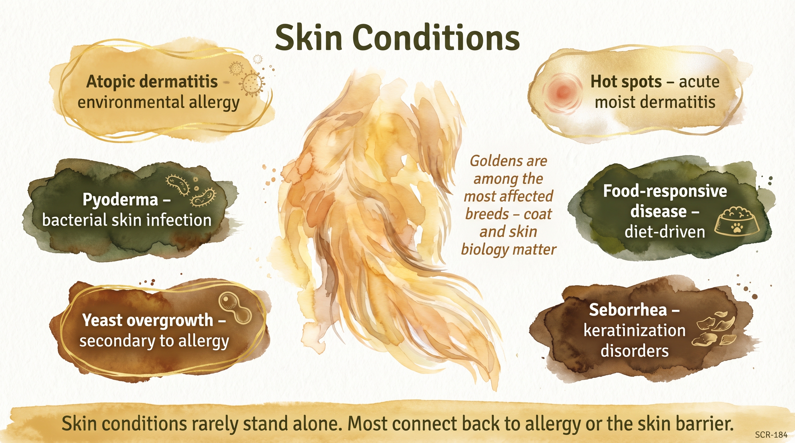
Skin conditions rarely stand alone - most connect back to allergy or the skin barrier.
Key Takeaways
- Most canine skin disease is not one simple thing. Allergy, barrier dysfunction, infection, and self-trauma often overlap.
- Goldens are a common chronic-skin-disease breed, especially for itch, hot spots, and allergy-linked problems.
- Recurrent skin infection usually means there is an underlying reason the skin keeps becoming vulnerable.
- Families help most by observing pattern, body sites, and recurrence rather than trying to guess the exact diagnosis from appearance alone.
The Evidence
This entry uses documented claim-level tags beyond the dedicated EvidenceBlocks below. These claims should remain tied to the entry Sources and SCR references during the next evidence-chain authoring pass.
- Canine dermatology consensus literaturedogs
Atopic dermatitis is the dominant chronic allergic skin disease in dogs, with secondary bacterial and yeast overgrowth commonly riding on top of barrier dysfunction. - Veterinary clinical dermatologydogs
Hot spots, pyoderma, and otitis often occur as downstream expressions of itch and self-trauma rather than as isolated primary diseases. - Diet-trial literaturedogs
Food-responsive skin disease requires properly structured elimination-diet methodology rather than casual food switching.
- Breed-predisposition literatureGolden Retrievers
Goldens are widely recognized as a common atopy and chronic otitis breed in practice, although the exact burden varies by dataset and referral pattern. - SCR-112 differential reminderGolden Retrievers
Ichthyosis remains a real Golden-specific skin differential and should not be conflated with ordinary allergy or infection.
- domestic dogs
No published study directly compares the most effective long-term management paths for skin conditions in dogs in dogs across breeds and ordinary home settings.
SCR References
Sources
- Hensel, P., Santoro, D., Favrot, C., Hill, P., & Griffin, C. (2015). Canine atopic dermatitis: Detailed guidelines for diagnosis and allergen identification. BMC Veterinary Research, 11, 196. https://doi.org/10.1186/s12917-015-0515-5
- Favrot, C., Steffan, J., Seewald, W., & Picco, F. (2010). A prospective study on the clinical features of chronic canine atopic dermatitis and its diagnosis. Veterinary Dermatology, 21(1), 23-31. https://doi.org/10.1111/j.1365-3164.2009.00758.x
- Olivry, T., Mueller, R. S., & Prelaud, P. (2015). Critically appraised topic on adverse food reactions of companion animals (1): Duration of elimination diets. BMC Veterinary Research, 11, 225. https://doi.org/10.1186/s12917-015-0541-3
- Mueller, R. S., Olivry, T., & Prelaud, P. (2016). Critically appraised topic on adverse food reactions of companion animals (2): Common food allergen sources in dogs and cats. BMC Veterinary Research, 12, 9. https://doi.org/10.1186/s12917-016-0633-8
- 2023 AAHA Management of Allergic Skin Diseases in Dogs and Cats Guidelines Task Force. (2023). 2023 AAHA management of allergic skin diseases in dogs and cats guidelines. Journal of the American Animal Hospital Association, 59(6), 255-284. https://doi.org/10.5326/JAAHA-MS-7396
- Bradley, C. W., Mauldin, E. A., & Morris, D. O. (2023). A review of cutaneous hypersensitivity reactions in dogs: A diagnostician's guide to allergy. Veterinary Pathology, 60(6), 839-852. https://doi.org/10.1177/03009858231189298