Mast Cell Tumors
Compound evidence detail2 SCRs / 5 parts
- Ambiguousany single percentage figure presented as a universal Golden Retriever cancer constant without naming the population, study design, or referral basis
- Documentedthe necropsy and referral cohort findings themselves; cancer as a leading cause of death in Golden Retrievers with honest sourcing and explicit dataset attribution
- Documentedthe Patnaik three-tier and Kiupel two-tier histologic grading systems, surgical margin and depth as independent prognostic variables, and grade as the dominant determinant of clinical behavior across the indolent-to-aggressive spectrum
- DocumentedGolden Retriever over-representation among breeds with elevated mast-cell-tumor incidence above the multi-breed canine baseline, supporting the clinical posture that any new cutaneous mass on a Golden warrants veterinary evaluation rather than watchful waiting
- Estimatedany specific numerical figure for Golden Retriever MCT incidence - source-dependent and not citable as a single universal breed constant
Mast cell tumors are one of the most frustrating canine cancers because they refuse to look consistent. Some appear as small, quiet skin lumps and are cured with surgery. Others behave aggressively, spread early, and require multimodal treatment. This variability is why veterinary oncologists sometimes call mast cell tumors "the great pretender" or "the great imitator." In practice, the lesson for families is simple: any new lump on a Golden Retriever deserves prompt evaluation rather than watchful optimism. Documented
What It Means
What Mast Cells Are
Mast cells are normal immune cells involved in allergy and inflammation. They contain granules rich in histamine and other mediators. When mast cells become neoplastic, they form tumors that can arise in the skin, subcutaneous tissues, or less commonly internal organs.
Because these are inflammatory cells, mast cell tumors can do two things at once behave like a mass; and trigger biologic effects from mediator release.
That is why some tumors become suddenly red, puffy, or itchy, and why some dogs develop gastrointestinal signs related to histamine release.
Why They Fool Families
Mast cell tumors do not have one standard look.
They may appear hairless or hairy; soft or firm; ulcerated or smooth; small and button-like; broad and plaque-like; and stable for a while and then suddenly inflamed. Documented
This visual inconsistency is the reason families get misled by the appearance of "just a bump." There is no safe visual rule that reliably distinguishes a harmless skin lump from a mast cell tumor.
Why Goldens Matter
Goldens are one of the breeds in which mast cell tumors are common enough to matter in everyday practice. That does not mean every skin lesion in a Golden is a mast cell tumor. It does mean the breed sits in the group where low-threshold lump evaluation is wise.
In the larger Golden cancer conversation, mast cell tumors matter because they add a different pattern than hemangiosarcoma or lymphoma. They are often more externally visible, more variable in behavior, and more dependent on grading and margins for prognosis. Observed-JB
How Diagnosis Happens
This is one of the cancers where diagnosis is often remarkably accessible.
Fine-needle aspirate is usually highly informative because mast cells have a characteristic cytologic appearance. A veterinarian can sample the mass quickly, often without sedation, and many tumors are provisionally identified the same day.
Once removed, the tumor is graded by histopathology. Two grading systems are most often discussed the older Patnaik system; and the newer two-tier Kiupel system.
The broad principle is that lower-grade tumors usually carry better outcomes, while high-grade tumors have higher metastatic and recurrence risk. Observed-JB
Why It Matters for Your Dog
Why Grade and Margins Matter So Much
Unlike some cancers where location or subtype dominates the story, mast cell tumor prognosis often hinges on pathology details histologic grade; mitotic index; margin status; tumor location; and nodal involvement. Observed-JB
A small low-grade cutaneous tumor removed completely can carry an excellent prognosis. A high-grade tumor, a tumor with incomplete margins, or a tumor in a more problematic location may require staging, repeat surgery, radiation, or systemic therapy. Documented
This is why families should resist two opposite mistakes assuming every mast cell tumor is mild; and assuming every mast cell tumor is catastrophic.
The pathology report is what clarifies the situation.
Treatment
Surgical excision is the cornerstone for most cutaneous mast cell tumors. The goal is complete removal with appropriate margins. Depending on pathology and staging, veterinarians may also discuss revision surgery; radiation therapy; chemotherapy; tyrosine kinase inhibitors; and supportive medications to manage histamine-related effects.
Again, the right framing is not "there is one mast cell treatment." The right framing is "treatment depends on grade, margins, spread, and location."
Prognosis
Prognosis ranges widely.
Low-grade tumors removed cleanly can have excellent long-term control. High-grade tumors or tumors with metastatic spread carry a much more guarded outlook. Subcutaneous mast cell tumors may behave differently from classic cutaneous ones, which is another reason careful pathology matters.
Families generally do better when the prognosis is not guessed from the look of the lump or from internet anecdotes. The pathology report and staging are the central tools here.
The Practical Family Rule
If you find a new lump on a Golden, make the appointment.
That advice is simple because the condition itself is not. "Watch it for a few months" is not ideal when one quick aspirate can often distinguish a tumor requiring action from something much less important.
This does not mean panic over every skin bump. It means replacing delay with information.
When to See a Veterinarian
Veterinary evaluation is warranted for any new skin or subcutaneous lump, a lump that changes size quickly, a mass that becomes red, swollen, itchy, or ulcerated, repeated vomiting or stomach upset in a dog with a known mast cell tumor, and enlarged local lymph nodes near a known skin mass.
The same-day emergency threshold is lower if the dog has a known mast cell tumor plus severe vomiting, weakness, collapse, or signs of gastrointestinal bleeding.
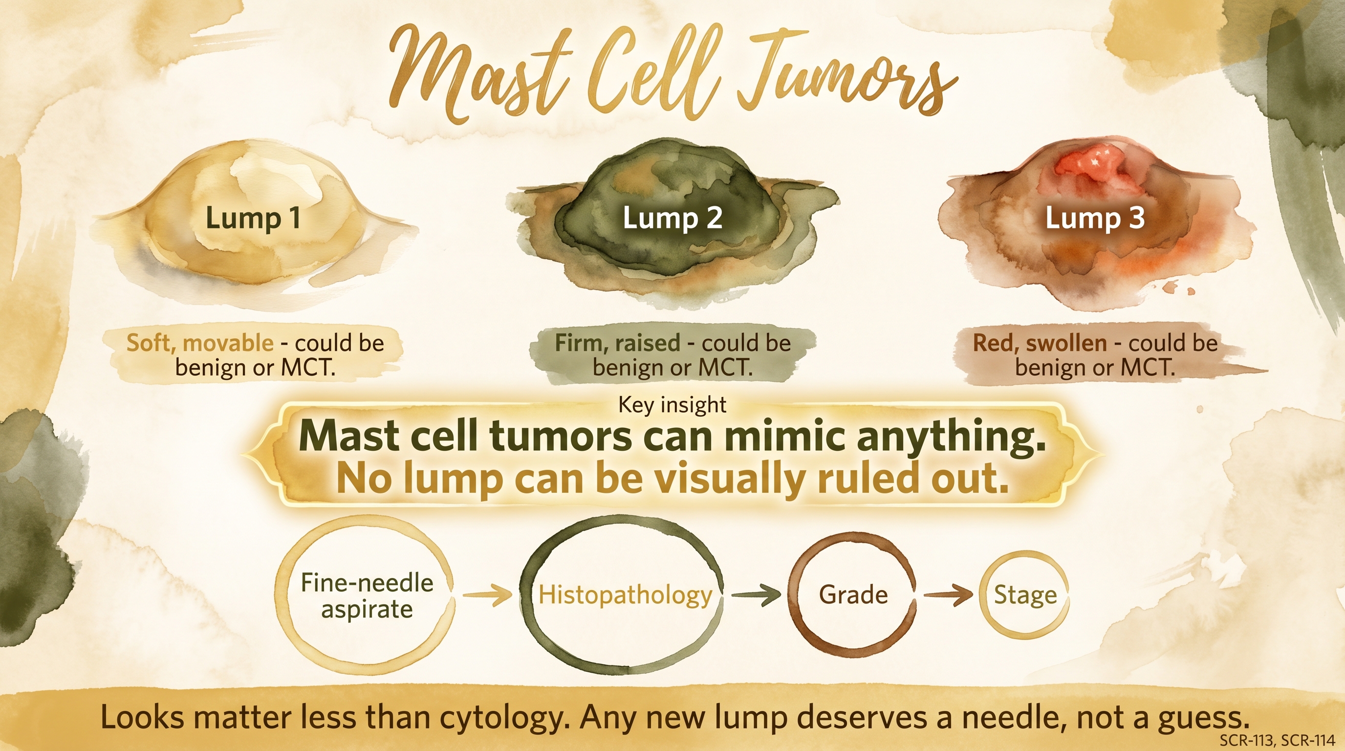
Looks matter less than cytology - any new lump deserves a needle, not a guess.
Key Takeaways
- Mast cell tumors are common in dogs and common enough in Goldens that any new lump deserves timely evaluation.
- These tumors are clinically variable, which means appearance alone is a poor guide to seriousness.
- Fine-needle aspirate is often diagnostic, and prognosis depends heavily on grade, margins, and spread.
- Some mast cell tumors are cured with surgery, while others require multimodal treatment, so pathology is central to honest counseling.
The Evidence
This entry uses observed claim-level tags beyond the dedicated EvidenceBlocks below. These tags mark JB program observation or practice-derived claims that need dedicated EvidenceBlock coverage in a later content pass.
- Veterinary oncology literaturedogs
Mast cell tumors are common canine skin tumors and fine-needle aspirate is usually highly diagnostic. - Patnaik and Kiupel grading literaturedogs
Histologic grade and margin status are major determinants of prognosis and treatment planning. - Golden cancer contextGolden Retrievers
Mast cell tumors are one of the major cancers contributing to Golden Retriever cancer burden.
- SCR placeholder noteGolden Retrievers
The SCR does not yet contain a dedicated mast-cell-tumor entry encoding the full grading and prognosis framework summarized here. - Breed-line heterogeneityGolden Retrievers
Breed predisposition is real, but line-level outcome prediction remains more complex than a simple one-number risk estimate.
SCR References
Sources
- Christensen, J., Johnson, K., Ettinger, S., Garrett, L., Gordon, I., Ireifej, S., Love, A., & Wisecup, M. (2026). 2026 AAHA oncology guidelines for dogs and cats. Journal of the American Animal Hospital Association, 62(1), 1-37. https://doi.org/10.5326/JAAHA-MS-7549
- Patnaik, A. K., Ehler, W. J., & MacEwen, E. G. (1984). Canine cutaneous mast cell tumor: Morphologic grading and survival time in 83 dogs. Veterinary Pathology, 21(5), 469-474. https://doi.org/10.1177/030098588402100503
- Kiupel, M., Webster, J. D., Bailey, K. L., Best, S., DeLay, J., Detrisac, C. J., Fitzgerald, S. D., Gamble, D., Ginn, P. E., Goldschmidt, M. H., Hendrick, M. J., Howerth, E. W., Janovitz, E. B., Langohr, I., Lenz, S. D., Lipscomb, T. P., Miller, M. A., Misdorp, W., Moroff, S., ... & Miller, R. (2011). Proposal of a 2-tier histologic grading system for canine cutaneous mast cell tumors to more accurately predict biological behavior. Veterinary Pathology, 48(1), 147-155. https://doi.org/10.1177/0300985810386469