Echocardiography in Dogs
Echocardiography is the test that turned canine cardiac screening from educated listening into direct structural and hemodynamic assessment. That shift matters enormously in Golden Retrievers because the breed's key inherited heart disease, subvalvular aortic stenosis, can be mild enough to evade simple auscultation while still being biologically real and breeding-relevant. In other words, the heart can have a problem before the stethoscope has a sound to hear. Documented
What It Means
What Echocardiography Is
Echocardiography is cardiac ultrasound. It uses sound waves to create moving images of the heart and, with Doppler techniques, to measure blood flow through valves and outflow tracts. Documented
In practical veterinary medicine, the exam can include two-dimensional imaging for anatomy, M-mode for chamber and wall measurements, color Doppler for flow direction and turbulence, pulsed-wave Doppler for measuring flow at specific sites, and continuous-wave Doppler for high-velocity jets. Documented
Families do not need to memorize those terms. They do need to understand that an echocardiogram is not just "an ultrasound picture." It is a structured cardiac exam that can reveal both anatomy and blood-flow behavior.
Why It Matters So Much in Goldens
Golden Retrievers are one of the breeds where screening methodology matters almost as much as the diagnosis itself. The inherited lesion of greatest concern is subvalvular aortic stenosis, a narrowing below the aortic valve that alters outflow.
Mild or subclinical cases can be breeding-relevant even when the dog looks normal and even when a murmur is absent or subtle. This is why the SCR now states the central screening conclusion plainly: specialist echocardiography is necessary for reliable SAS screening.
That statement is not anti-stethoscope. A stethoscope is still useful. It is just not enough for the load-bearing breeding question.
What the Exam Can Show
A proper echocardiogram can show chamber size, wall thickness, valve motion, outflow-tract structure, turbulence and velocity patterns, and consequences of pressure overload or abnormal flow. Documented
For SAS specifically, Doppler matters because it quantifies the velocity of blood as it moves through the left ventricular outflow tract and across the aortic valve region. That is a much stronger screening tool than depending on whether a sound is audible in a given room on a given day.
Why Doppler Changes the Game
Without Doppler, much of the screening question would still depend on indirect interpretation. Doppler adds measurable hemodynamics.
That matters because blood-flow velocity can reveal obstruction severity; turbulence can be visualized rather than inferred; borderline cases can be interpreted with more structure; and serial screening becomes more comparable over time.
This is why breeder language like "heart checked" is often too vague to be useful. A casual primary-care auscultation and a board-certified cardiology echo are not interchangeable events.
Why It Matters for Your Dog
OFA Cardiac Context
Echocardiography also matters because families often encounter cardiac screening through the OFA ecosystem. In that setting, it helps to know that cardiac entries may sit on top of different exam depths.
Broadly speaking, the strongest clearance architecture is evaluation by a board-certified cardiologist; Doppler echocardiography rather than auscultation alone; and serial reassessment when the result is equivocal or the breeding decision is consequential.
The practical lesson is simple: registry language is only as strong as the method under it.
Why Specialist Training Matters
Cardiology is one of the clearest fields where operator expertise matters. Image acquisition, probe positioning, Doppler angle, and interpretation all affect the meaning of the exam.
A cardiologist is not only better equipped to run the test. A cardiologist is better equipped to decide whether a finding is truly abnormal; whether a velocity is meaningful or stress-influenced; whether a repeat study is needed; and how a borderline finding should affect breeding decisions. Documented
That matters because false reassurance is one of the major risks in screening. Families and breeders are often much more aware of false positives than of false negatives. For SAS, false reassurance is the more dangerous problem.
The Limits of Echocardiography
Even very good screening has limits.
Important caveats include screening does not guarantee lifelong freedom from later disease, mild lesions can be challenging at the margins, physiologic stress can influence Doppler readings, and a single normal exam does not eliminate the value of serial screening in some dogs. Documented
This does not weaken the case for echo. It clarifies how to use echo honestly.
Why This Page Matters for Families
Most puppy buyers are not choosing whether to schedule an echo themselves for breeding stock. They are deciding whether the breeder's health-testing language is meaningful.
This page helps them ask the right questions was the cardiac screening done by a cardiologist; was Doppler echocardiography performed; was the result entered in a public registry; and if the result was borderline, was the dog rechecked.
Those questions are far more useful than simply asking whether the parents were "heart cleared."
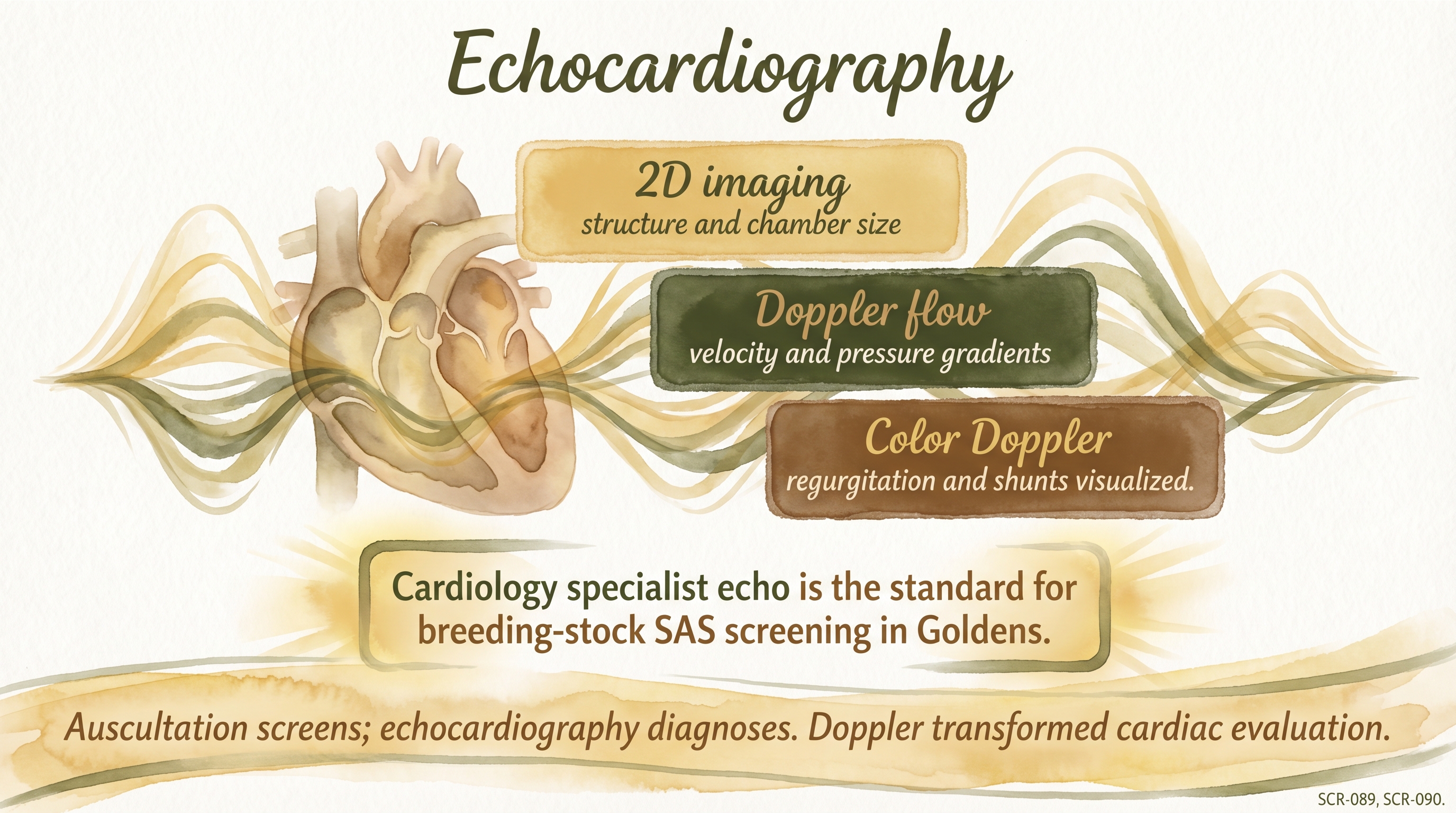
Auscultation screens; echocardiography diagnoses. Doppler transformed cardiac evaluation.
Key Takeaways
- Echocardiography is cardiac ultrasound plus flow measurement, not just a moving picture of the heart.
- In Golden Retrievers, specialist Doppler echo is the gold-standard screening method for SAS because auscultation alone can miss important disease.
- Cardiac registry labels are only as meaningful as the method and expertise underneath them.
- A normal echo is highly valuable, but good screening still respects borderline findings, serial follow-up, and the limits of any one exam.
The Evidence
- SCR-061 supportdogs and Golden Retrievers
Reliable SAS screening requires specialist echocardiography because auscultation alone misses clinically relevant and breeding-relevant disease. - Veterinary cardiology literaturedogs
Doppler methods make cardiac screening meaningfully stronger by measuring blood-flow velocity and turbulence rather than relying on sound alone. - OFA cardiac-screening contextdogs
Registry language around heart clearance is only as informative as the exam depth and examiner expertise behind it.
- Clinical cardiology practicedogs
Borderline or stress-sensitive findings sometimes require serial follow-up rather than one definitive claim from a single screening session. - Breeding-screening logicdogs
The purpose of screening is risk reduction and better selection, not a guarantee that no cardiac disease can ever appear later in the dog or offspring.
- domestic dogs
No published study directly defines the single best testing interval, threshold, or decision rule for echocardiography in dogs across all Golden Retriever households and breeding programs.
SCR References
Sources
- Orthopedic Foundation for Animals. (n.d.). Cardiac disease. https://ofa.org/diseases/other-diseases/cardiac-disease/
- Orthopedic Foundation for Animals. (2023). Overview of recent changes to the OFA cardiac clearances. https://ofa.org/wp-content/uploads/2023/01/OFA-ACA-Press-Release-12-22-2022.pdf
- Stern, J. A., Meurs, K. M., Nelson, O. L., Lahmers, S. M., & Lehmkuhl, L. B. (2012). Familial subvalvular aortic stenosis in Golden Retrievers: Inheritance and echocardiographic findings. Journal of Small Animal Practice, 53(4), 213-216. https://doi.org/10.1111/j.1748-5827.2011.01187.x
- Ontiveros, E. S., Fousse, S. L., Crofton, A. E., Hodge, T. E., Gunther-Harrington, C. T., Visser, L. C., & Stern, J. A. (2019). Congenital cardiac outflow tract abnormalities in dogs: Prevalence and pattern of inheritance from 2008 to 2017. Frontiers in Veterinary Science, 6, 52. https://doi.org/10.3389/fvets.2019.00052
- van Staveren, M. D. B., & Szatmari, V. (2020). Detecting and recording cardiac murmurs in clinically healthy puppies in first opinion veterinary practice at the first health check. Acta Veterinaria Scandinavica, 62, 57. https://doi.org/10.1186/s13028-020-00535-1